What's New?
Globe denotes a link or links that goes offsite from VHA Diagnostic Electron Microscopy. You may wish to review the privacy notices on those sites since their information collection practices may differ from ours.
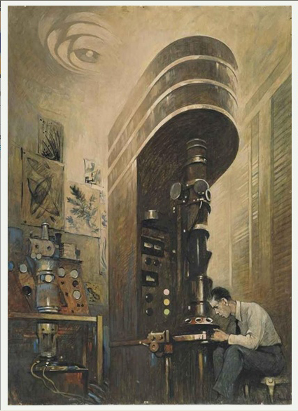
Art Captures Science as an electron microscope and operator are interpreted in the Industrial Machine Age style from post-World War II 1940's. Titled 'The Electron Microscope', by artist Thornton Oatley, this watercolor, gouache, and charcoal painting was commissioned by the National Geographic Society and published in National Geographic Magazine, December, 1945, p. 747, Figure XI, with the caption "Electron Microscope Opens Even the Secrets of the Molecule." The physical principles of electrons and lenses in Electron Microscopes parallel those of photons and lenses in Light Microscopes, but magnifications of over a million times are feasible. 
![]() Dr. Bezhad Najafian, Associate Professor of Pathology and Director, Electron Microscopy, Department of Pathology, University of Washington, Seattle, presented his studies of Quantitative Electron Microscopy in Fabry Nephropathy as part of the Benchmarks for Quality in Ultrastructual Pathology series . If you missed this excellent presentation, link to the recording at
Dr. Bezhad Najafian, Associate Professor of Pathology and Director, Electron Microscopy, Department of Pathology, University of Washington, Seattle, presented his studies of Quantitative Electron Microscopy in Fabry Nephropathy as part of the Benchmarks for Quality in Ultrastructual Pathology series . If you missed this excellent presentation, link to the recording at http://va-eerc-ees.adobeconnect.com/py11pznl4jt5/.
Contact VHA National EM Program QA/QM Coordinator Ann LeFurgey, Edoris.LeFurgey@va.gov, to be placed on the mailing list for future programs in the 'Benchmarks' series.
Highlights of the Benchmarks for Quality in Ultrastructural Pathology Live Meeting Series
Dr. Sergey Brodsky, Ohio State University, Columbus, Ohio, discussed multiple aspects of Anticoagulant-Related Nephropathy: Clinical and Pathology Considerations as it relates to ultrastructure, nephrology and clinical presentation and treatment. If you missed this excellent presentation, link to the recording at http://va-eerc-ees.adobeconnect.com/py11pznl4jt5/.
The Role of Electron Microscopy in the Diagnosis and Causation of Asbestos-Related Diseases Among US Veterans was broadcast by pulmonary diseases expert Dr. Victor Roggli, Department of Pathology, Duke University Medical Center, Durham, NC. This presentation included a discussion of the role of electron microscopy in the diagnosis and causation of asbestos-related diseases among US Veterans. A link to the recording of the webinar is now available:
Mark Haas, MD, PhD, Cedars Sinai Medical Center, Los Angeles, CA, spoke in detail about his work on the Value of Electron Microscopy in Evaluation of Renal Allograft Biopsies. The audio/video capture of this detailed presentation with outstanding images is available at http://va-eerc-ees.adobeconnect.com/p30jdwas3m6/.
Cynthia Goldsmith, CDC/OID/NCEZID, Atlanta, GA, presented a timely webinar on viruses such as Zika, Chikungunya, Ebola and case studies describing the clinical and environmental settings in which they occur in March, 2016. VHA EES has provided a link to its excellent audio/video recording of this webinar at http://va-eerc-ees.adobeconnect.com/p4tr8bcdqa2/.
Karen Weidenheim, MD, Albert Einstein College of Medicine, Montefiore Medical Center, Bronx, NY, gave an excellent presentation on Successful Handling of Skeletal Muscle Biopsies in the General Pathology and Electron Microscopy Laboratories. An audio/video recording of her talk is now available through VHA-EES at http://va-eerc-ees.adobeconnect.com/p81b2tjtzfi/. Dr. Weidenheim defined the role of the pathologist in the diagnosis of muscle disorders such as muscular dystrophy, rare genetic myopathies, congenital and toxic myopathies, with emphasis on employing correlative methodologies from ultrastructural morphology to molecular genetics.
![]() The FY2021 annual activity report and cases for peer review for VHA Diagnostic EM are due on December 5, 2021 and will be reviewed by the Ad Hoc Peer Review Committee in early spring 2022. The VISN, Medical Center and Facility Director will receive the annual summary review letter in July, 2022. The Diagnostic EM SharePoint site is available for electronic submission of reports, data and electron micrographs. EM facility directors have been pre-registered for access to the site. Support staff who may be uploading reports or images can also be given access to the site. Feedback and comments welcomed. Contact EM QA/QM Coordinator for access or questions (Edoris.LeFurgey@va.gov).
The FY2021 annual activity report and cases for peer review for VHA Diagnostic EM are due on December 5, 2021 and will be reviewed by the Ad Hoc Peer Review Committee in early spring 2022. The VISN, Medical Center and Facility Director will receive the annual summary review letter in July, 2022. The Diagnostic EM SharePoint site is available for electronic submission of reports, data and electron micrographs. EM facility directors have been pre-registered for access to the site. Support staff who may be uploading reports or images can also be given access to the site. Feedback and comments welcomed. Contact EM QA/QM Coordinator for access or questions (Edoris.LeFurgey@va.gov).
 The Microscopy Society of America (MSA) Certification Board has announced dates for 2022 Certified Electron Microscopy Technologist (CEMT) Exams. Individuals with the requisite educational and/or occupational qualifications can attain certification by completing an application and passing both written and practical examinations. Successful candidates are awarded an official numbered MSA Certificate and a Certification Pin. Two examination cycles are offered each year.
For more information and exam schedule, contact CEMT. Electron Microscopy technical training programs are valuable staffing resources for VA EM programs seeking to fill vacancies. The
San Joaquin Delta College Electron Microscopy Program, Stockton, CA, also offers EM certification and an associate arts degree for students.
 The 2021 VHA National Diagnostic EM Program Ad Hoc Review Group Meeting and Workshop is tentatively planned in conjunction with the 111th Annual Meeting of the USCAP in Los Angeles, CA, March 19-24, 2022, including the companion meeting of the Society for Ultrastructural Pathology, to be held on Sunday, 20 March, 4:30-6:30 pm. Dr. Guillermo Herrera, member of the VA EM Ad Hoc Committee, will present an Ultrastructural Evaluation of Immune Mediated Renal Injury at the Companion Meeting. The VA EM Ad Hoc members' workshop will be devoted to in depth review of FY21 diagnostic cases submitted as part of the annual peer evaluation, as well as evaluation of the newly revised VHA P&LMS Directive incorporating Diagnostic Electron Microscopy and long term plans for the VA Diagnostic EM Program.
![]()
Microscopy Meetings, Conferences and Workshops- Ultrapath XX, the biennial meeting of the
Society for Ultrastructural Pathology, was postponed in 2020 and 2021 due to the pandemic travel restrictions. The Ultrapath XX meeting is now tentatively scheduled again in Amsterdam, Holland, for June, 2022 (dates to be determined). These meetings focus exclusively on application of electron microscopy in pathologic diagnosis and research of human and animal diseases. For more information and the most recent details, see
ultrapath.microscopie.nl. [The program of the last meeting in 2018 is available on the Society website (
www.ultrapath.org).] Microscopy and Microanalysis, the annual meeting of the Microscopy Society of America (
http://www.microscopy.org) will take place in Portland, Oregon, from July 31-August 4, 2022. The Diagnostic and Biomedical Microscopy Focused Interest Group (FIG) members will hold several sessions, titled Challenges and Advances in Electron Microscopy for Research and Diagnosis of Diseases in Human, Plants and Animals; FIG contact is Claudia Lopez (lopezcl@ohsu.edu). Information and calendars about other current EM meetings and conferences are available from the Microscopy Society of America.
Equivalent of Green Fluorescent Protein for Electron Microscopy? The development of a small, highly engineered plant (Arabidopsis thaliana) protein, dubbed "miniSOG," may elevate the abilities of electron microscopy in the same way that green fluorescent protein (GFP) and its relatives have made modern light microscopy in biological research much more powerful and useful ( PLoS Biol 9(4): e1001041. doi:10.1371/journal.pbio.1001041).
CDC - Electron Microscopy for Rapid Diagnosis of Infectious Agents in Emergent Situations
EM Retention requirements are on pages 73-74 of VHA HANDBOOK 1106.01 (Jan. 29, 2016)*. A full copy of the Handbook is available on the VA Intranet Diagnostic EM SharePoint Site or the national P&LMS website.
*See example memorandum for appropriate turn-in of EM documents, blocks, slides exceeding retention times.
Check out the TEM cases page. John G. Guccion, M.D., EM Director of the Washington DC Program, contributed a set of twelve interesting cases for your review. In addition a series of SEM/TEM and x-ray microanalysis cases submitted by the Durham EM Program are now available.
The Society for Ultrastructural Pathology publishes a series of Newsletters regarding electron microscopy and ultrastructural pathology on their web site -
Ultrastructural Pathology Newsletters.
Generic Electron Microscopy processing procedures for Kidney biopsy, Tissue and Nerve biopsy are available for review and possible modification for local use. To see examples of generic processing procedures for EM Kidney biopsy, Tissue biopsy and Nerve biopsy click on the underlined links. As other generic procedures are added they will be available on the Generic Procedures page where the above biopsy specimen procedures are listed.
Some CAP inspectors may interpret that Diagnostic EM Programs should have external evaluations of their cases twice a year, rather than the annual review. This question was presented to the CAP accreditation division by Michael Brophy, former National Enforcement Officer. See the question and response by CAP (CAP question).
Information on setting up sharing agreements - Need to bring in some additional money to support your EM Program? See the Sharing agreement section for more details.
Please share your good ideas and suggestions with other EM facilities. If you have any ideas or recommendations to share which save time, resources or effort in producing diagnostic EM reports for our patients, please send that information to us for possible addition to this web page. Potential examples include microwave processing procedures, EM specimen submission information/criteria or forms for use by referring facilities, etc.
Please e-mail Edoris.LeFurgey@va.gov with your comments and suggestions.


