
History of VA and its administrations
What is the history of the Department of Veteran Affairs? The origin story of the modern VA includes several previous entities and names dating as far back as the American colonies. To help understand that legacy, this site serves as the centralized digital location for VA’s history with the focus of educating about the special relationship between the nation and its Veterans, including how that has impacted society. Here there are various stories and exhibits that examine the individuals, institutions and innovations of the last 250 years. There have been multiple agencies that have provided the service and benefits to America’s Veterans, including the Bureau of Pensions, National Home for Disabled Volunteer Soldiers, National Cemetery System, Bureau of War Risk Insurance, Veterans Bureau, and Veterans Administration, all of which are referenced on the preceding pages.
Eventually, VA’s story will be displayed to the public at the National VA History Center at the Dayton, Ohio VA campus. All information related to that effort will be found here as well.
This site is updated regularly, so check back often or sign up for our monthly newsletter in the sign up box below to have the latest stories in your inbox.
Latest VA History Posts
Featured Stories
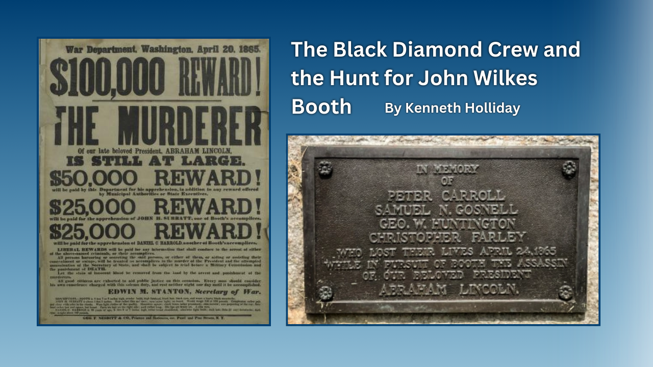
During the late evening, early hours of April 23-24, 1865, the Black Diamond, a ship on the Potomac River searching for President Abraham Lincoln's assassin John Wilkes Booth collided with another ship, the USS Massachusetts. The incident was a terrible accident during the frantic mission to locate the fleeing Booth before he escaped into Virginia. Unfortunately many lives were lost, including four civilians who had been summoned from a local fire department by the Army. For their assistance during this military operation, all four were buried in the Alexandria National Cemetery, some of the few civilians to receive that honor.
History of VA in 100 Objects
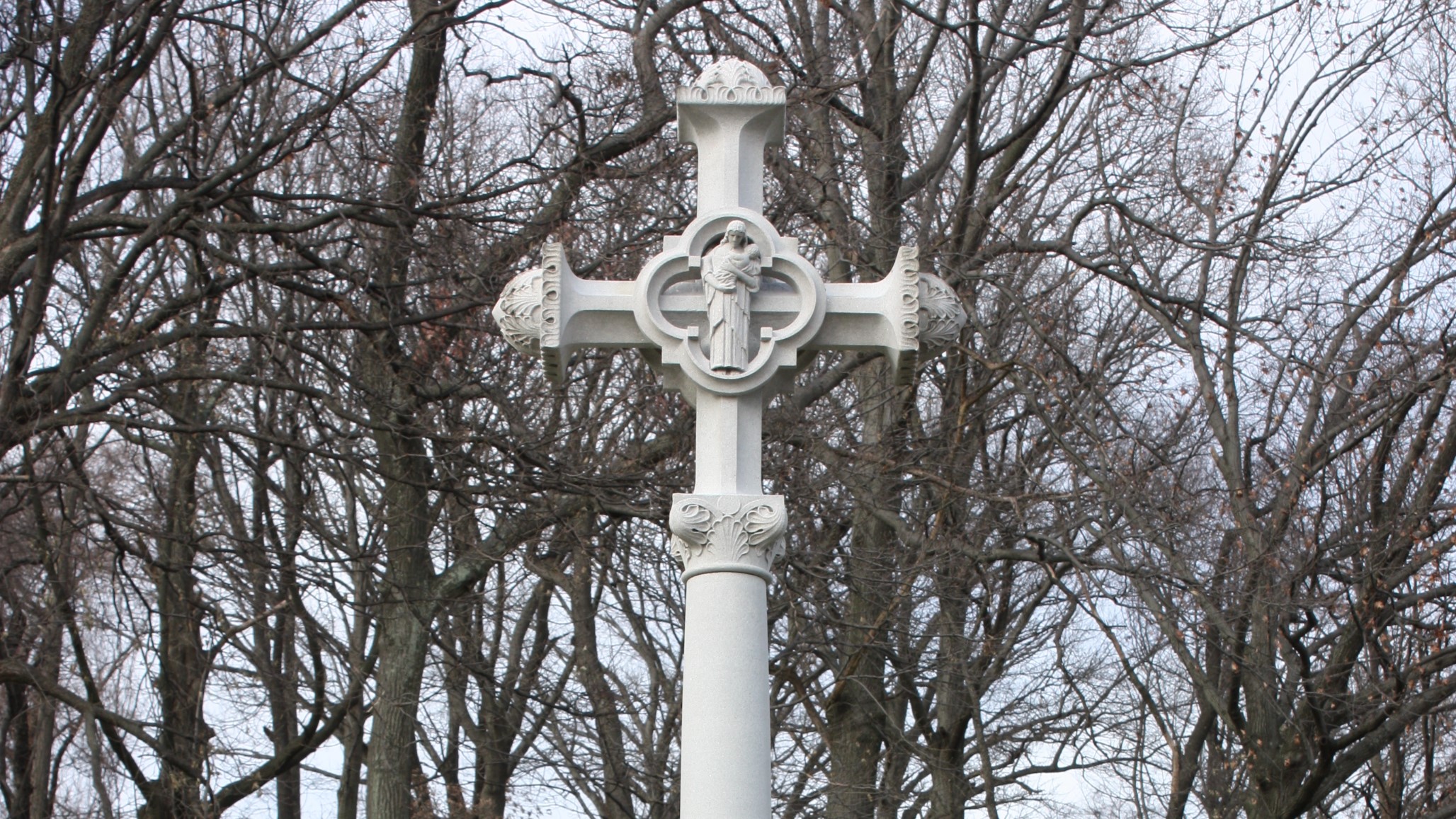
In the waning days of World War I, French sailors from three visiting allied warships marched through New York in a Liberty Loan Parade. The timing was unfortunate as the second wave of the influenza pandemic was spreading in the U.S. By January, 25 of French sailors died from the virus.
These men were later buried at the Cypress Hills National Cemetery and later a 12-foot granite cross monument, the French Cross, was dedicated in 1920 on Armistice Day. This event later influenced changes to burial laws that opened up availability of allied service members and U.S. citizens who served in foreign armies in the war against Germany and Austrian empires.
Curator Corner
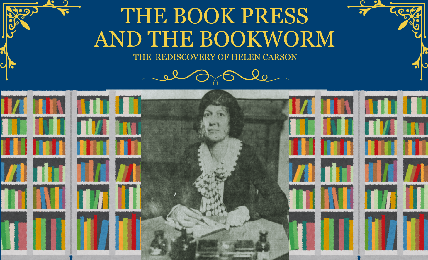
It isn’t often that researchers who work with historic objects get to know the people who used those objects every day. Sometimes we get lucky and can link artifacts to certain facilities or buildings on a historic VA campus, but usually we must look for more hidden lines of evidence to figure out how an object fits into the history of those who care for our Nation’s Veterans. As nice as it would be, it isn’t as if many artifacts turn up labeled with their owners’ names! So, imagine my surprise when my teammates and I began sweeping Putnam Library for any historic objects left behind before the building is closed for renovation, and found just that.
As far as artifacts go, its story seemed simple: book presses like these would have been used to help maintain and repair the thousands of books read in Putnam Library ever since it first opened in 1879. The day that I first got up close and personal with the press, I noticed a woman’s name scraped into the black paint of the platen (the technical name for the big metal plate used to hold books together). It said “Helen Carson” in big, legible letters. As we carefully transported the heavy press down the many stairs inside Putnam Library, I looked at the name and thought “Hm…wonder who that is?”.
History of VA in 100 Objects
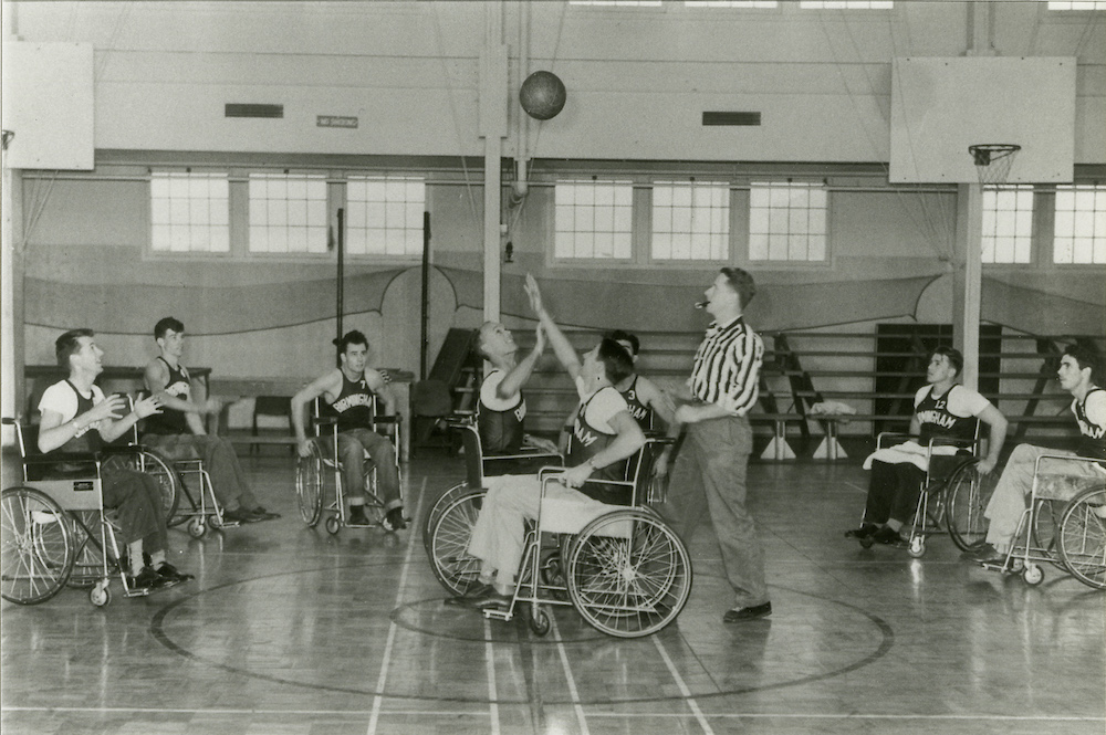
Basketball is one of the most popular sports in the nation. However, for paraplegic Veterans after World War II it was impossible with the current equipment and wheelchairs at the time. While VA offered these Veterans a healthy dose of physical and occupational therapy as well as vocational training, patients craved something more. They wanted to return to the sports, like basketball, that they had grown up playing. Their wheelchairs, which were incredibly bulky and commonly weighed over 100 pounds limited play.
However, the revolutionary wheelchair design created in the late 1930s solved that problem. Their chairs featured lightweight aircraft tubing, rear wheels that were easy to propel, and front casters for pivoting. Weighing in at around 45 pounds, the sleek wheelchairs were ideal for sports, especially basketball with its smooth and flat playing surface. The mobility of paraplegic Veterans drastically increased as they mastered the use of the chair, and they soon began to roll themselves into VA hospital gyms to shoot baskets and play pickup games.
Exhibits
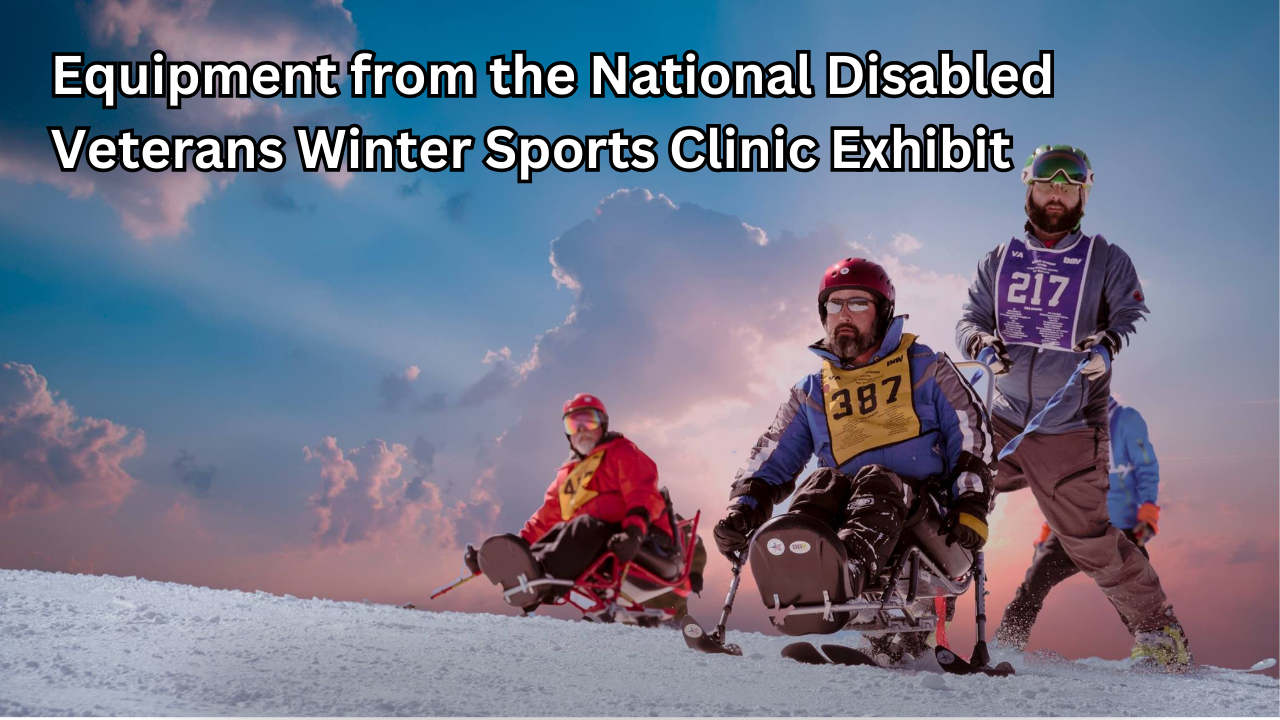
In 2024, the National Disabled Veterans Winter Sports Clinic, hosted by the Grand Junction VA Medical Center and co-presented with Disabled American Veterans, will donate adaptive ski equipment from the early years of the event to the National VA History Center in Dayton, Ohio. The clinic started in 1986 when VA established it and then held the the inaugural event at Powderhorn the following year. The clinic welcomed approximately 90 Veterans from 27 states, bolstered by a volunteer staff of about 20. Despite skepticism from many health professionals of the era, the clinic underscored the importance of physical activity for persons of all abilities.
This exhibit includes multiple items that were used at the early clinics, which are finding a new home at the National VA History Center.
History of VA in 100 Objects
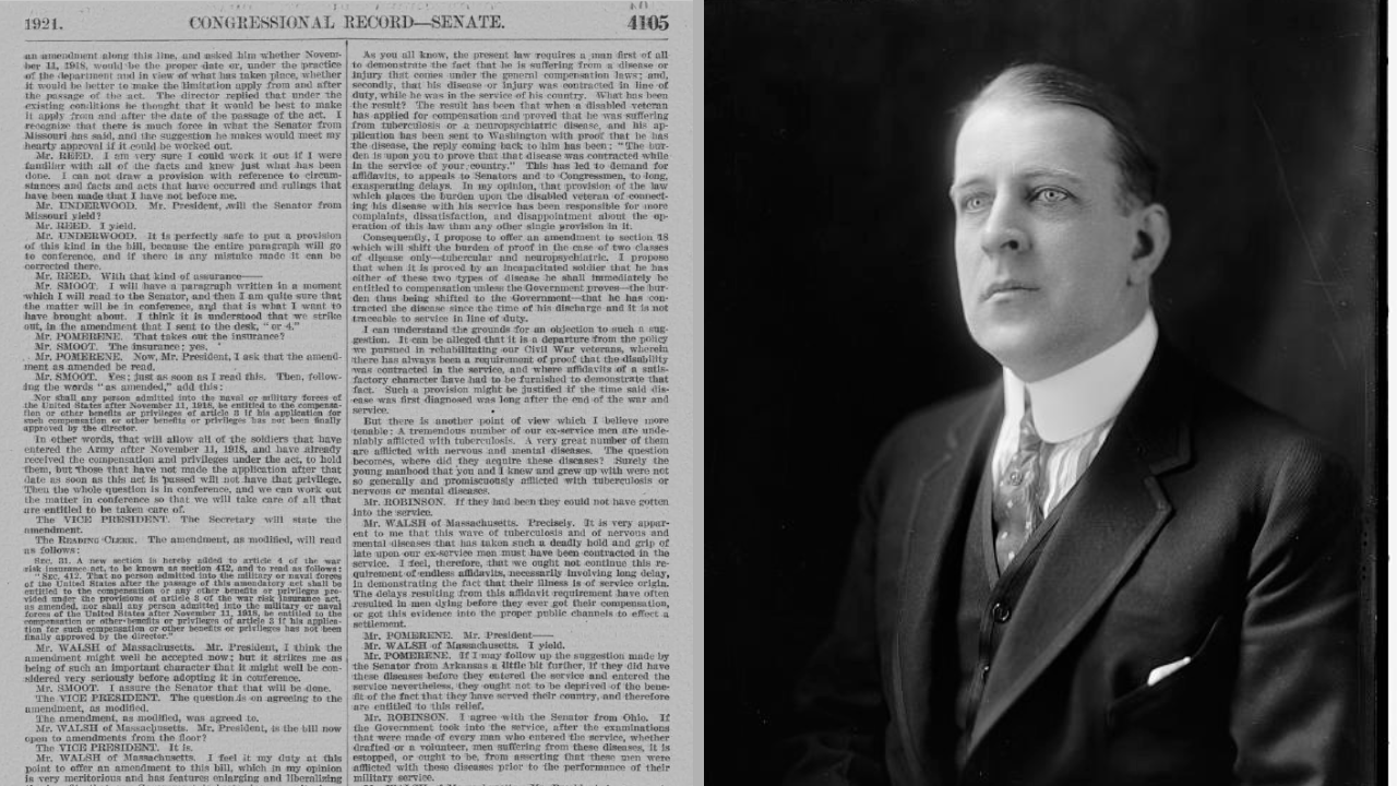
After World War I, claims for disability from discharged soldiers poured into the offices of the Bureau of War Risk Insurance, the federal agency responsible for evaluating them. By mid-1921, the bureau had awarded some amount of compensation to 337,000 Veterans. But another 258,000 had been denied benefits. Some of the men turned away were suffering from tuberculosis or neuropsychiatric disorders. These Veterans were often rebuffed not because bureau officials doubted the validity or seriousness of their ailments, but for a different reason: they could not prove their conditions were service connected.
Due to the delayed nature of the diseases, which could appear after service was completed, Massachusetts Senator David Walsh and VSOs pursued legislation to assist Veterans with their claims. Eventually this led to the first presumptive conditions for Veteran benefits.
National VA History Center
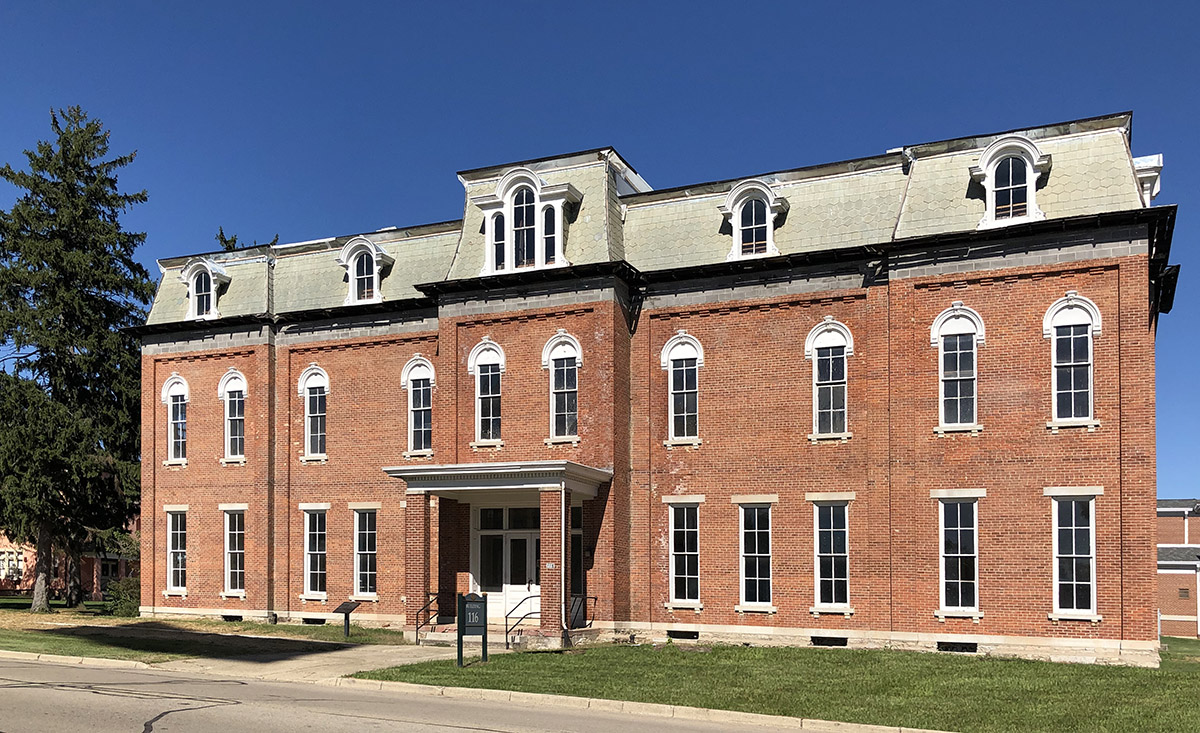
The National VA History Center is the forthcoming museum and archival center for the historical collection and records pertaining to the Department of Veterans Affairs and its legacy agencies. It is located at the Dayton VA Medical Center campus, itself a designated National Historic Landmark. While the idea of the history center is not new, recent actions to make it a reality are. Ceremonies marking its official establishment – and the start of renovation work on two historic buildings to house the collection – were held in August 2020. The public opening of the facility isn’t expected for several years, but great work is ongoing behind the scenes. Look for monthly updates in the Curator Corner, illustrating the multi-faceted steps that go into establishing a museum and its collection as well as the archives with its growing amount of records from scratch. This will be the GO-TO home for the History Center.
History of VA in 100 Objects exhibit
The History of VA in 100 Objects exhibit spotlights the objects that illuminate how the nation has honored and cared for Veterans from 1776 to the present. New entries are added every few weeks as we continue the countdown to the 100th Object. Click on the link below to view all of the entries published to date or you can search them by using the magnifying glass icon on this page. Join the journey through VA’s past, object by object.
History of VA in 100 Objects
In the waning days of World War I, French sailors from three visiting allied warships marched through New York in a Liberty Loan Parade. The timing was unfortunate as the second wave of the influenza pandemic was spreading in the U.S. By January, 25 of French sailors died from the virus.
These men were later buried at the Cypress Hills National Cemetery and later a 12-foot granite cross monument, the French Cross, was dedicated in 1920 on Armistice Day. This event later influenced changes to burial laws that opened up availability of allied service members and U.S. citizens who served in foreign armies in the war against Germany and Austrian empires.
History of VA in 100 Objects
Basketball is one of the most popular sports in the nation. However, for paraplegic Veterans after World War II it was impossible with the current equipment and wheelchairs at the time. While VA offered these Veterans a healthy dose of physical and occupational therapy as well as vocational training, patients craved something more. They wanted to return to the sports, like basketball, that they had grown up playing. Their wheelchairs, which were incredibly bulky and commonly weighed over 100 pounds limited play.
However, the revolutionary wheelchair design created in the late 1930s solved that problem. Their chairs featured lightweight aircraft tubing, rear wheels that were easy to propel, and front casters for pivoting. Weighing in at around 45 pounds, the sleek wheelchairs were ideal for sports, especially basketball with its smooth and flat playing surface. The mobility of paraplegic Veterans drastically increased as they mastered the use of the chair, and they soon began to roll themselves into VA hospital gyms to shoot baskets and play pickup games.
Featured Stories
Our Featured Stories section provides scholarly researched and written content on the people, places, events and innovations that illustrate VA’s remarkable evolution serving Veterans since the start of our nation. These are longer stories, with historical details on the rich lineage of VA and the significant moments in time.
Featured Stories
During the late evening, early hours of April 23-24, 1865, the Black Diamond, a ship on the Potomac River searching for President Abraham Lincoln's assassin John Wilkes Booth collided with another ship, the USS Massachusetts. The incident was a terrible accident during the frantic mission to locate the fleeing Booth before he escaped into Virginia. Unfortunately many lives were lost, including four civilians who had been summoned from a local fire department by the Army. For their assistance during this military operation, all four were buried in the Alexandria National Cemetery, some of the few civilians to receive that honor.
Featured Stories
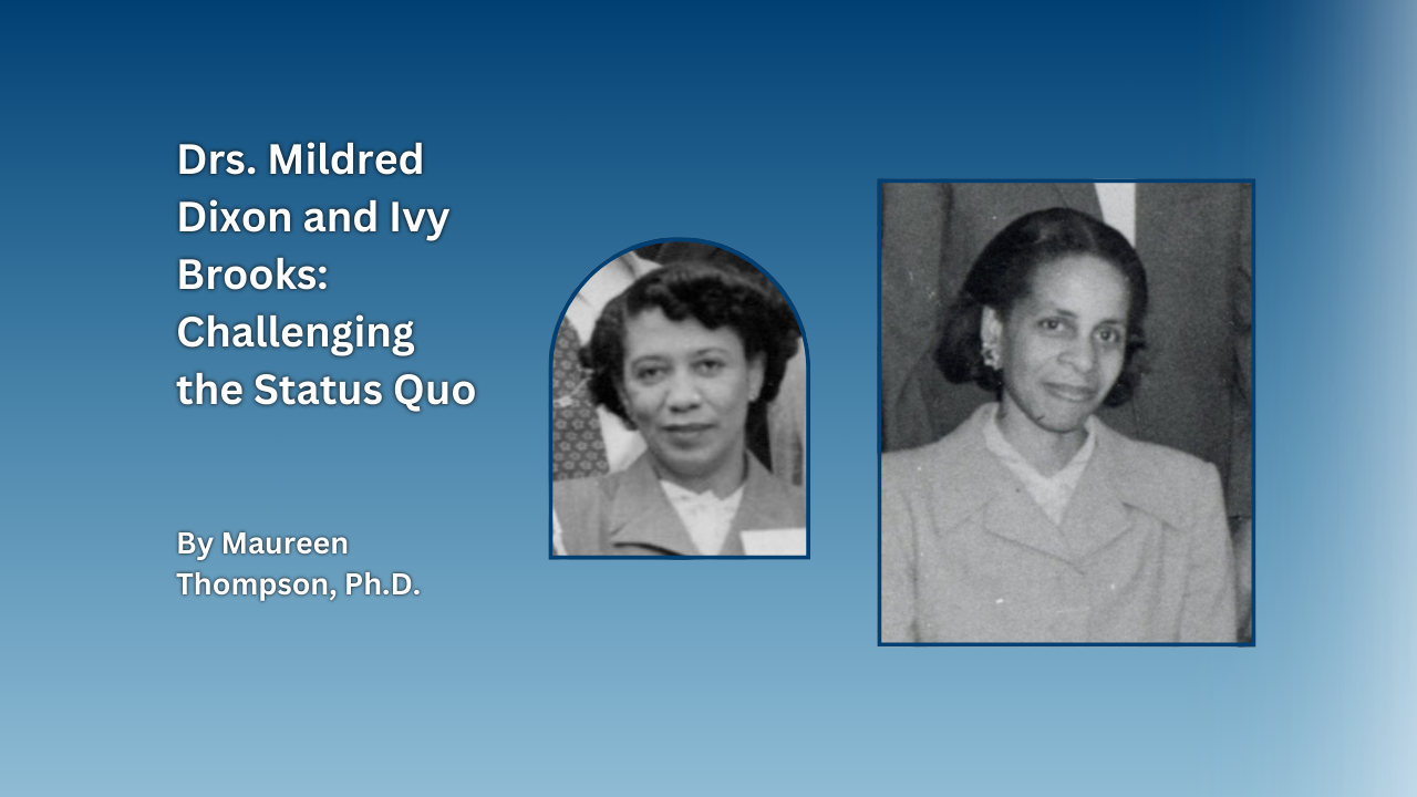
In the mid-twentieth century, the lives of Dr. Ivy Brooks and Mildred Dixon, two trailblazing Black women physicians, converged at the Tuskegee, Alabama, VA Medical Center. Doctor's Ivy Roach Brooks and Mildred Kelly Dixon shared much in common. Both women were born in 1916 in the northeastern United States and received training in East Orange, New Jersey. They both launched careers in alternate medical professions before entering the fields of radiology and podiatry, respectively. Pioneering many “firsts” throughout their professional lives, both women faced and overcame the rampant racism and sexism of the era.
Virtual Exhibits
A collection of virtual exhibits covering various historical aspects of VA, presented with focus on visual images that have been pulled from archives across the nation. Most use an exhibit-host to showcase specific topics with compelling pictures and research that go beyond stories and features.
Exhibits
In 2024, the National Disabled Veterans Winter Sports Clinic, hosted by the Grand Junction VA Medical Center and co-presented with Disabled American Veterans, will donate adaptive ski equipment from the early years of the event to the National VA History Center in Dayton, Ohio. The clinic started in 1986 when VA established it and then held the the inaugural event at Powderhorn the following year. The clinic welcomed approximately 90 Veterans from 27 states, bolstered by a volunteer staff of about 20. Despite skepticism from many health professionals of the era, the clinic underscored the importance of physical activity for persons of all abilities.
This exhibit includes multiple items that were used at the early clinics, which are finding a new home at the National VA History Center.
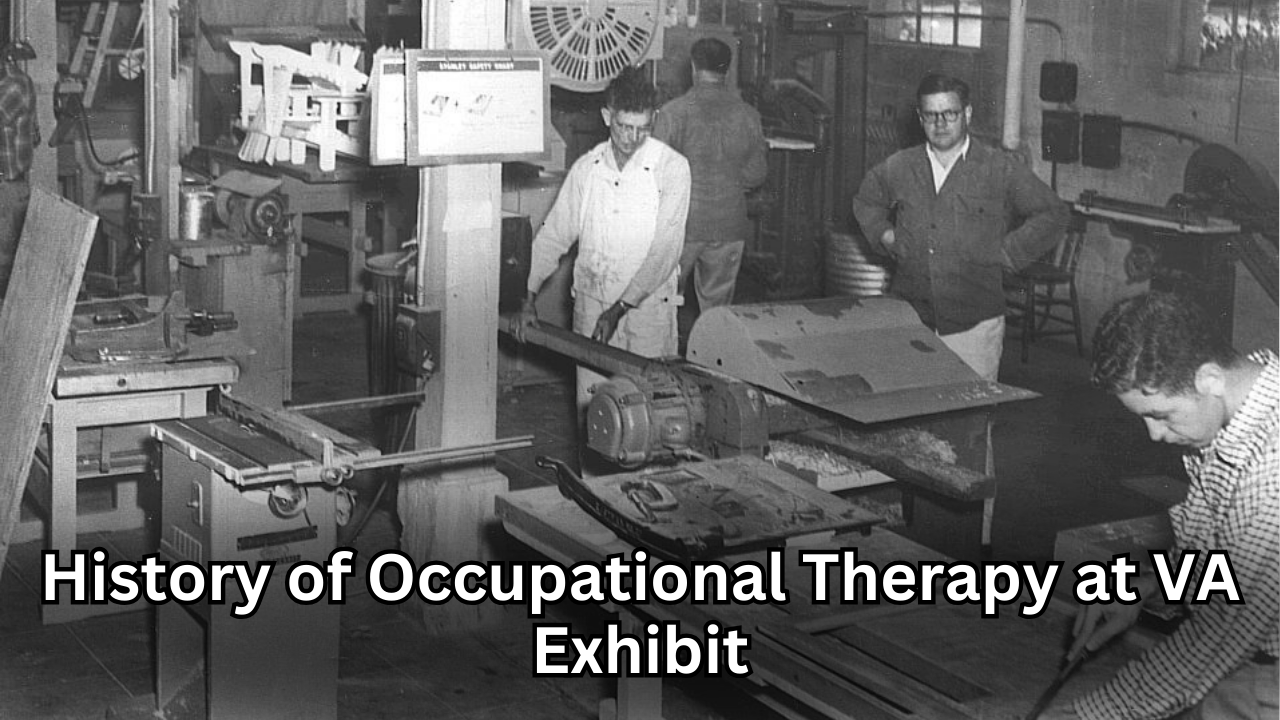
Exhibits
While Veterans engaged in activities and learned trades at the National Home for Disabled Volunteer Soldiers (NHDVS) since its inception after the Civil War, formal occupational therapy programs became components of rehabilitative care for Veterans beginning in the 20th century. This exhibit explores what type of activities were used to treat Veterans by showing items from the collection at the National VA History Center.
Curator Corner
While the National VA History Center continues progressing, get a peek inside the growing artifact collection and records archive that will one day be featured in various displays or be accessible and centralized for research. As part of our effort, the Curator Kurt Senn and Senior Archivist Robyn Rodgers will provide insight into the efforts behind the scenes to establish the future museum and archive center that will help show VA’s story. Some of our series include ‘What’s in the box?‘ that will highlight unique items that are literally found in boxes as the team opens them and ‘From the collection,’ which traces the history of items that are stored on site.
Curator Corner
It isn’t often that researchers who work with historic objects get to know the people who used those objects every day. Sometimes we get lucky and can link artifacts to certain facilities or buildings on a historic VA campus, but usually we must look for more hidden lines of evidence to figure out how an object fits into the history of those who care for our Nation’s Veterans. As nice as it would be, it isn’t as if many artifacts turn up labeled with their owners’ names! So, imagine my surprise when my teammates and I began sweeping Putnam Library for any historic objects left behind before the building is closed for renovation, and found just that.
As far as artifacts go, its story seemed simple: book presses like these would have been used to help maintain and repair the thousands of books read in Putnam Library ever since it first opened in 1879. The day that I first got up close and personal with the press, I noticed a woman’s name scraped into the black paint of the platen (the technical name for the big metal plate used to hold books together). It said “Helen Carson” in big, legible letters. As we carefully transported the heavy press down the many stairs inside Putnam Library, I looked at the name and thought “Hm…wonder who that is?”.
Curator Corner
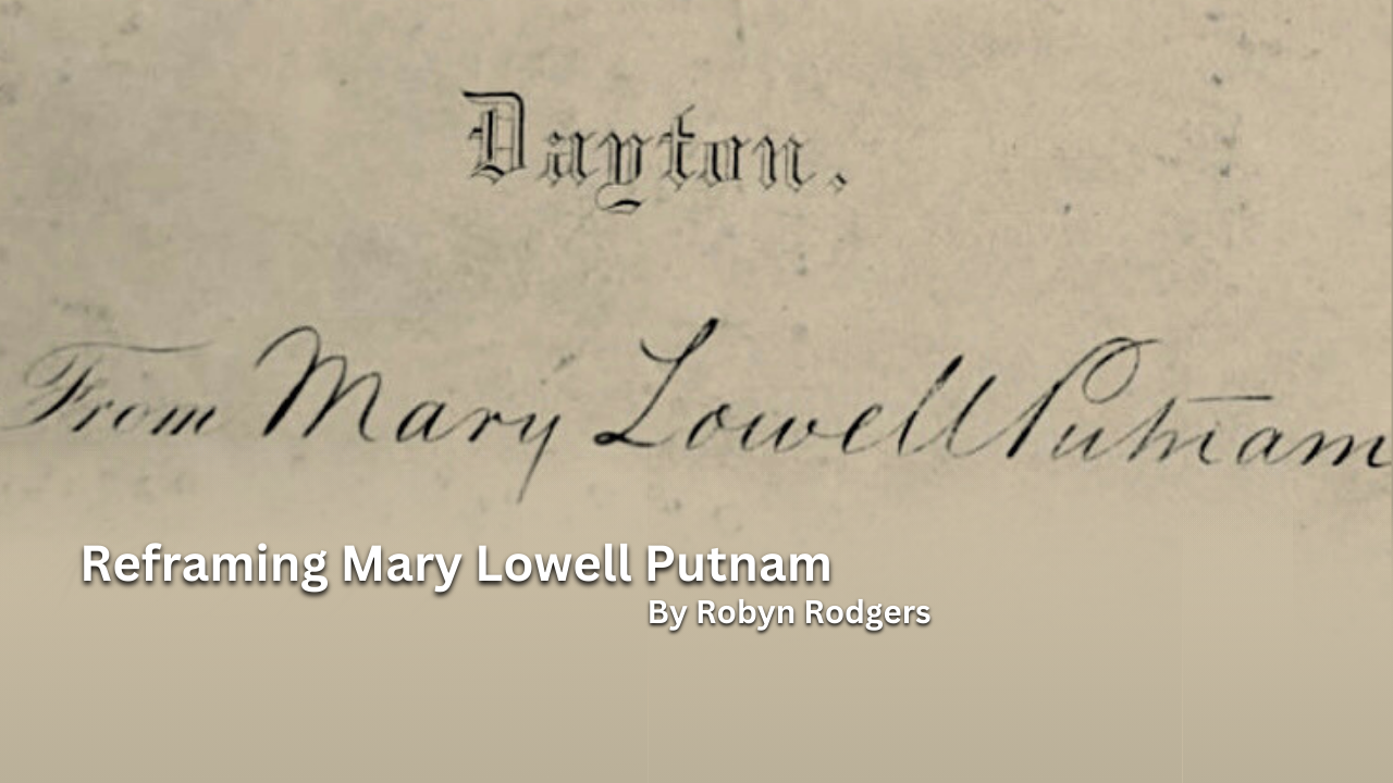
Mary Lowell Putnam is tied to VA history by her generous donation of a large volume of books to the Central Branch of the National Home for Disabled Volunteer Soldiers. These books, meant to honor her son who died in the Civil War, helped foster reading advancement for the Veterans who lived there after the war and into the 20th Century. However, her life was more than just a moment in time donating books. It included a life-long study of languages and a very sharp opinion that she shared in writing throughout her life.

